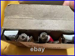
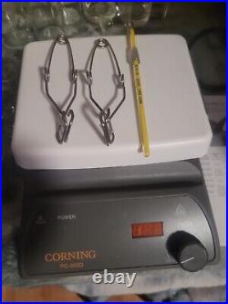

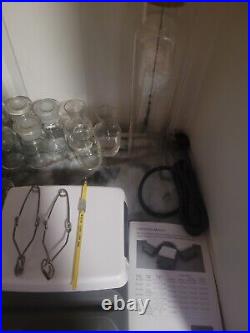

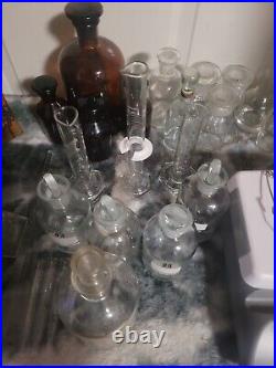
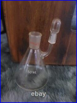
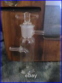
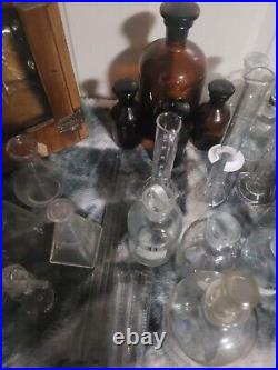
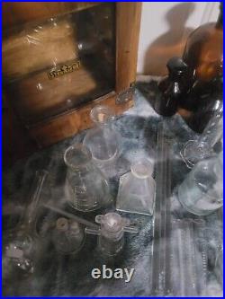
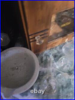

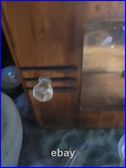




Large Lot Chemistry Lab Equipment & Supplies new, great condition or vintage. Everything is in excellent condition many brand new items. Some vintage, some great shape, some new. List of items pictured. Cornings 8 heat plate PC-400D New. Five piece beaker set 50-1000ml units New. Granite mortar no pestle. Large flask with cork. Brown lab flask set with glass stoppers. Pyrex 50ml cylinders new. Pyrex short 50ml cylinders. Flask with glass stoppers. Glass flask with glass stopper. Lab glass device 2 outlets and glass stopper. Glass Lab device with outlet. Pyrex 250ml glass Lab device with outlet. Vintage sterizer wooden and glass face excellent condition from the 1950s. Everything list has no chips or cracks and is in great shape new items will come in the original box. This item is in the category “Business & Industrial\Healthcare, Lab & Dental\Medical, Lab & Dental Supplies\Glassware & Plasticware\Other Medical & Lab Glassware”. The seller is “manchesterpawn2008″ and is located in this country: US. This item can be shipped to United States.
- Brand: PYREX
- Model: varios lot
- MPN: 1212
- Country/Region of Manufacture: United States
- Intended Use/Discipline: Biological Laboratory, Physical Laboratory, Medical Laboratory
Incoming search terms:
- https://vintagemedicalequipment com/tag/chemistry/
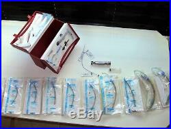




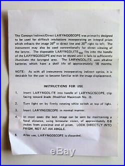
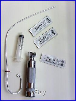
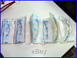

Portex Emergency Intubation Kit, 1968, Vintage Medical Equipment, Great Condition. Emergency Airway Kit designed for the Difficult Airway. This kit contains 2 laryngoscope handles(one pediatric and one adult), an assortment of oral airways, a metal stylet, one metal laryngoscope handle, and a series of endotracheal tubes from infant to large adult. Enlarge and read the pic with the page of instructions. This kit contains a prism built into the laryngoscope that can be used to visualize airway openings that are unable to detect with normal direct laryngoscopy. The prism allows 30 degrees of refraction in-line, and 20 degrees of refraction left-to-right. The prism is unique to me. I have never seen a difficult airway aid such as this. This kit is in excellent shape and would be a great addition to a collection of vintage medical equipment. The item “Portex Emergency Intubation Kit, 1968, Vintage Medical Equipment, Great Condition” is in sale since Monday, March 4, 2019. This item is in the category “Business & Industrial\Healthcare, Lab & Dental\Other Healthcare, Lab & Dental”. The seller is “sjc2m4″ and is located in Fredericksburg, Texas. This item can be shipped to United States.
- Brand: Portex
- Intended Use/Discipline: Anesthesiology
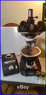
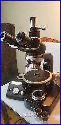
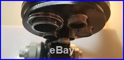
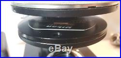
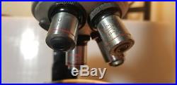
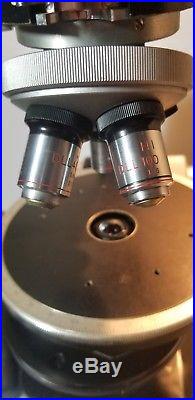
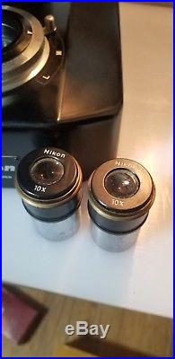

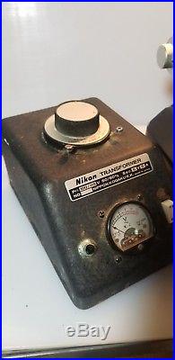
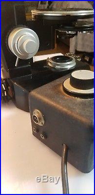
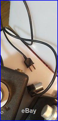
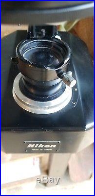
Microscope is in fantastic, working condition. Shows only minor wear from use, comes with an extra set of olympus optics. The bulb blew, so will need a replacement bulb. Transformer unfortunately has some rust, but works well. Also has a nikon interference phase contrast condenser. Plug end of bulb is too narrow to fit the power supply, so assuming the cord is not original to this unit. Units were tested separately of each other. Excellent, rare example of a great microscope. What you see in pictures is what you will receive. The item “Nikon S-ke S L-ke microscope, 1960s vintage scope, extra optics, works great” is in sale since Monday, November 19, 2018. This item is in the category “Business & Industrial\Healthcare, Lab & Dental\Medical & Lab Equipment, Devices\Microscopes”. The seller is “ytmarshall” and is located in Culpeper, Virginia. This item can be shipped to United States, Canada, United Kingdom, Denmark, Romania, Slovakia, Bulgaria, Czech republic, Finland, Hungary, Latvia, Lithuania, Malta, Estonia, Australia, Greece, Portugal, Cyprus, Slovenia, Japan, China, Sweden, South Korea, Indonesia, Taiwan, Thailand, Belgium, France, Hong Kong, Ireland, Netherlands, Poland, Spain, Italy, Germany, Austria, Bahamas, Israel, Mexico, New Zealand, Philippines, Singapore, Switzerland, Norway, Saudi arabia, Ukraine, United arab emirates, Qatar, Kuwait, Bahrain, Croatia, Malaysia, Chile, Colombia, Costa rica, Panama, Trinidad and tobago, Guatemala, Honduras, Jamaica, Antigua and barbuda, Aruba, Belize, Dominica, Grenada, Saint kitts and nevis, Saint lucia, Montserrat, Turks and caicos islands, Barbados, Bangladesh, Bermuda, Brunei darussalam, Bolivia, Egypt, French guiana, Guernsey, Gibraltar, Guadeloupe, Iceland, Jersey, Jordan, Cambodia, Cayman islands, Liechtenstein, Sri lanka, Luxembourg, Monaco, Macao, Martinique, Maldives, Nicaragua, Oman, Pakistan, Paraguay, Reunion.
- Brand: Nikon
- Model: S-ke
- Optical/Digital: Optical
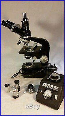
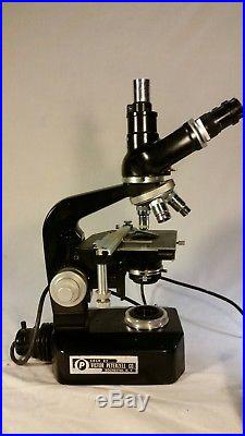
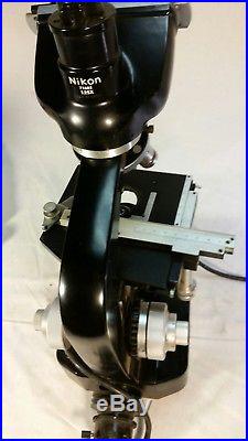
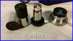
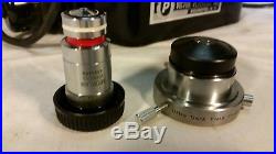
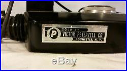
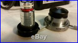
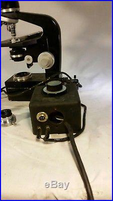
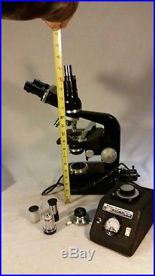
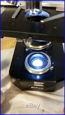
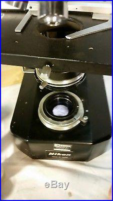
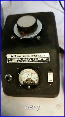
Rare Nikon s-ke microscope in great working condition with Kohler illuminator, transformer and extra optics. Nikon 71482 1.25x labeled on upper back. Everything works great and is in excellent condition. Sale includes everything in pictures. I tested the microscope to the best of my ability and found it easy to use and that all dials and adjustments moved smoothly. The only thing i found with this Nikon S-ke set up that didn’t work as expected is the indicator light on the transformer. The transformer does work well and the dial on it moved as i adjusted the light. Please ask for any additional tests, pictures or additional information. I try and display and describe everything i sell honestly and correctly, taking care to note any and all imperfections. I hope this microscope find a new home that can utilize it’s potential and appreciate it’s form and function. Model S-Ke Microscope (Circa late 1960s) The S-Ke was the first Nikon microscope equipped with a Köhler illuminator built into the microscope base, enabling convenient and perfect photomicrography when a camera was mounted and used with one of Nikon’s Microflex adapters. A reflecting mirror was provided as an accessory and could replace the illumination field lens to provide light from a source outside the microscope. Like the basic Model S, interchangeable parts made the Model S-Ke microscope a versatile tool for studies in biology, medicine, and metallurgy. Depending on the user’s needs, it could be equipped with various combinations of eyepieces, eyepiece tubes, objectives, condensers, and stages. This microscope, built during the 1960s and 1970s, was provided with a coaxial set of coarse and fine focusing knobs, which were located near the base. A preset device allowed quick refocusing after the stage was lowered for changing a specimen or applying immersion oil. A wooden cabinet was also available for storage when the microscope was not in use. There were five selections of eyepiece tubes available: inclined monocular, inclined monocular with an inclined phototube, binocular, and two versions of trinocular. Four interchangeable stages provided a choice of surfaces for examining specimens: a rectangular mechanical stage, circular floating, graduated, circular rotatable, and a plain square stage. A variety of achromatic and apochromatic objectives were available, designed specifically for either metallurgical (episcopic) or biological/medical (diascopic) uses. Reviews on the Nikon s-ke From the point of view of design and functionality, it may be the pinnacle of the light microscope. It combines superb optics, smoothness of movement, a steady platform, and a beautiful streamlined design reminiscent of aerodynamic designs of the 1940′s. Since the era of the SKe, microscopes have become lighter and larger, more complex and complete, but not necessarily better. This particular model has a built in light and separate transformer that are capable of koehler illumination. It has concentric coarse and fine focus knobs with a stop lever to prevent the lens from lowering into the slide. The two binocular 10x eyepieces are also Nikon. According to Nikon; this Ke base houses a complete Koehler illuminating and optical system, making the most efficient use of available light, as only the area under inspection is illuminated. Glare, flare and halo effects are reduced to a minimum. A built-in base turret with auxiliary condenser lenses to adjust the illumination for low, medium and high power objectives is one of the features that makes it an ideal microscope for both visual and photomicrographic applications. About the ultra dark field condenser Darkfield contrast technique where only light diffracted from the specimen is used to form the image TECHNOLOGY: Darkfield microscopy creates contrast in transparent unstained specimens such as living cells. It depends on controlling specimen illumination so that central light which normally passes through and around the specimen is blocked. Rather than light illuminating the sample with a full cone of light (as in brightfield microscopy) the condenser forms a hollow cone with light travelling around the cone rather than through it. This form of illumination allows only oblique rays of light to strike the specimen on the microscope stage and the image is formed by rays of light scattered by the sample and captured in the objective lens. When there is no sample on the microscope stage the view is completely dark. Care should be taken in preparing specimens as features above and below the plane of focus can also scatter light and compromise image quality (for example, dust, fingerprints). In general, thin specimens are better because the possibility of diffraction artifacts is reduced. APPLICATIONS: In darkfield microscopy, contrast is created by a bright specimen on a dark background. It is ideal for revealing outlines, edges, boundaries, and refractive index gradients but does not provide a great deal of information about internal structure. Ideal subjects include living, unstained cells (where darkfield illumination provides information not visible with other techniques), although fixed stains cells can also be imaged successfully. Darkfield imaging is particularly useful in haematology for the examination of fresh blood. Non-biological specimens include minerals, chemical crystals, colloidal particles, inclusions and porosity in glass, ceramics, and polymer thin sections…. Objectives designed to be used with reflected darkfield illumination have a special construction consisting of a 360-degree hollow chamber surrounding the centrally located lens elements. Light from the illuminator passes through the periphery of the objective and is directed at the specimen from every azimuth in oblique rays to form a hollow cone of illumination. This is often accomplished by means of circular mirrors or prisms located at the bottom of the objective’s hollow chamber. In this manner, the objective serves as two separate optical systems coupled coaxially such that the outer system functions as a “condenser” and the inner system as a typical objective. The item “Nikon S-ke microscope, rare 1960s vintage scope, extra optics, works great” is in sale since Wednesday, July 25, 2018. This item is in the category “Business & Industrial\Healthcare, Lab & Life Science\Lab Equipment\Microscopes”. The seller is “konga33″ and is located in New Tazewell, Tennessee. This item can be shipped worldwide.
- Brand: Nikon
- Model: S-ke
- Optical/Digital: Optical
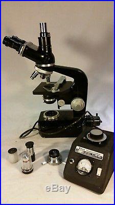
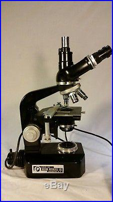
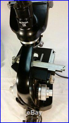
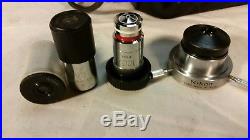
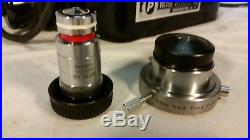
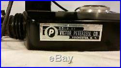
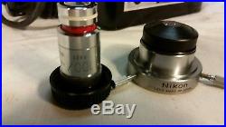
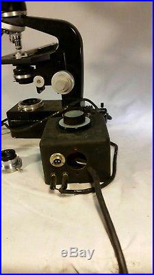
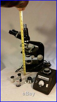
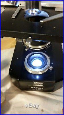
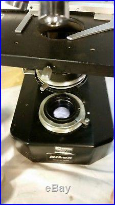
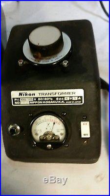
Rare Nikon s-ke microscope in great working condition with Kohler illuminator, transformer and extra optics. Nikon 71482 1.25x labeled on upper back. Everything works great and is in excellent condition. Sale includes everything in pictures. I tested the microscope to the best of my ability and found it easy to use and that all dials and adjustments moved smoothly. The only thing i found with this Nikon S-ke set up that didn’t work as expected is the indicator light on the transformer. The transformer does work well and the dial on it moved as i adjusted the light. Please ask for any additional tests, pictures or additional information. I try and display and describe everything i sell honestly and correctly, taking care to note any and all imperfections. I hope this microscope find a new home that can utilize it’s potential and appreciate it’s form and function. Model S-Ke Microscope (Circa late 1960s) The S-Ke was the first Nikon microscope equipped with a Köhler illuminator built into the microscope base, enabling convenient and perfect photomicrography when a camera was mounted and used with one of Nikon’s Microflex adapters. A reflecting mirror was provided as an accessory and could replace the illumination field lens to provide light from a source outside the microscope. Like the basic Model S, interchangeable parts made the Model S-Ke microscope a versatile tool for studies in biology, medicine, and metallurgy. Depending on the user’s needs, it could be equipped with various combinations of eyepieces, eyepiece tubes, objectives, condensers, and stages. This microscope, built during the 1960s and 1970s, was provided with a coaxial set of coarse and fine focusing knobs, which were located near the base. A preset device allowed quick refocusing after the stage was lowered for changing a specimen or applying immersion oil. A wooden cabinet was also available for storage when the microscope was not in use. There were five selections of eyepiece tubes available: inclined monocular, inclined monocular with an inclined phototube, binocular, and two versions of trinocular. Four interchangeable stages provided a choice of surfaces for examining specimens: a rectangular mechanical stage, circular floating, graduated, circular rotatable, and a plain square stage. A variety of achromatic and apochromatic objectives were available, designed specifically for either metallurgical (episcopic) or biological/medical (diascopic) uses. Reviews on the Nikon s-ke From the point of view of design and functionality, it may be the pinnacle of the light microscope. It combines superb optics, smoothness of movement, a steady platform, and a beautiful streamlined design reminiscent of aerodynamic designs of the 1940′s. Since the era of the SKe, microscopes have become lighter and larger, more complex and complete, but not necessarily better. This particular model has a built in light and separate transformer that are capable of koehler illumination. It has concentric coarse and fine focus knobs with a stop lever to prevent the lens from lowering into the slide. The two binocular 10x eyepieces are also Nikon. According to Nikon; this Ke base houses a complete Koehler illuminating and optical system, making the most efficient use of available light, as only the area under inspection is illuminated. Glare, flare and halo effects are reduced to a minimum. A built-in base turret with auxiliary condenser lenses to adjust the illumination for low, medium and high power objectives is one of the features that makes it an ideal microscope for both visual and photomicrographic applications. About the ultra dark field condenser Darkfield contrast technique where only light diffracted from the specimen is used to form the image TECHNOLOGY: Darkfield microscopy creates contrast in transparent unstained specimens such as living cells. It depends on controlling specimen illumination so that central light which normally passes through and around the specimen is blocked. Rather than light illuminating the sample with a full cone of light (as in brightfield microscopy) the condenser forms a hollow cone with light travelling around the cone rather than through it. This form of illumination allows only oblique rays of light to strike the specimen on the microscope stage and the image is formed by rays of light scattered by the sample and captured in the objective lens. When there is no sample on the microscope stage the view is completely dark. Care should be taken in preparing specimens as features above and below the plane of focus can also scatter light and compromise image quality (for example, dust, fingerprints). In general, thin specimens are better because the possibility of diffraction artifacts is reduced. APPLICATIONS: In darkfield microscopy, contrast is created by a bright specimen on a dark background. It is ideal for revealing outlines, edges, boundaries, and refractive index gradients but does not provide a great deal of information about internal structure. Ideal subjects include living, unstained cells (where darkfield illumination provides information not visible with other techniques), although fixed stains cells can also be imaged successfully. Darkfield imaging is particularly useful in haematology for the examination of fresh blood. Non-biological specimens include minerals, chemical crystals, colloidal particles, inclusions and porosity in glass, ceramics, and polymer thin sections…. Objectives designed to be used with reflected darkfield illumination have a special construction consisting of a 360-degree hollow chamber surrounding the centrally located lens elements. Light from the illuminator passes through the periphery of the objective and is directed at the specimen from every azimuth in oblique rays to form a hollow cone of illumination. This is often accomplished by means of circular mirrors or prisms located at the bottom of the objective’s hollow chamber. In this manner, the objective serves as two separate optical systems coupled coaxially such that the outer system functions as a “condenser” and the inner system as a typical objective. The item “Nikon S-ke microscope, rare 1960s vintage scope, extra optics, works great” is in sale since Saturday, June 23, 2018. This item is in the category “Business & Industrial\Healthcare, Lab & Life Science\Lab Equipment\Microscopes”. The seller is “konga33″ and is located in New Tazewell, Tennessee. This item can be shipped worldwide.
- Model: S-ke
- Brand: Nikon
- Optical/Digital: Optical
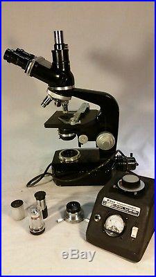
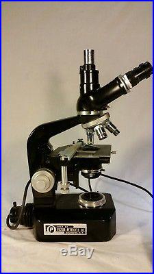
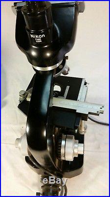
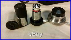
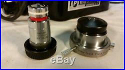
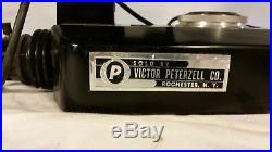
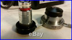
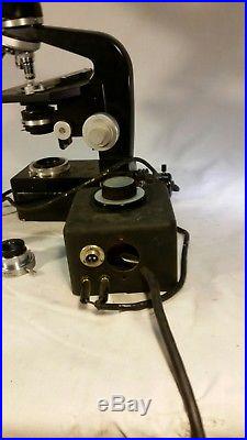
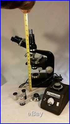
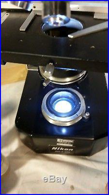
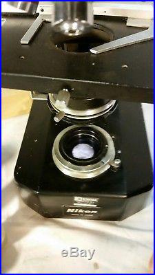
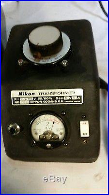
Rare Nikon s-ke microscope in great working condition with Kohler illuminator, transformer and extra optics. Nikon 71482 1.25x labeled on upper back. Everything works great and is in excellent condition. Sale includes everything in pictures. I tested the microscope to the best of my ability and found it easy to use and that all dials and adjustments moved smoothly. The only thing i found with this Nikon S-ke set up that didn’t work as expected is the indicator light on the transformer. The transformer does work well and the dial on it moved as i adjusted the light. Please ask for any additional tests, pictures or additional information. I try and display and describe everything i sell honestly and correctly, taking care to note any and all imperfections. I hope this microscope find a new home that can utilize it’s potential and appreciate it’s form and function. Model S-Ke Microscope (Circa late 1960s) The S-Ke was the first Nikon microscope equipped with a Köhler illuminator built into the microscope base, enabling convenient and perfect photomicrography when a camera was mounted and used with one of Nikon’s Microflex adapters. A reflecting mirror was provided as an accessory and could replace the illumination field lens to provide light from a source outside the microscope. Like the basic Model S, interchangeable parts made the Model S-Ke microscope a versatile tool for studies in biology, medicine, and metallurgy. Depending on the user’s needs, it could be equipped with various combinations of eyepieces, eyepiece tubes, objectives, condensers, and stages. This microscope, built during the 1960s and 1970s, was provided with a coaxial set of coarse and fine focusing knobs, which were located near the base. A preset device allowed quick refocusing after the stage was lowered for changing a specimen or applying immersion oil. A wooden cabinet was also available for storage when the microscope was not in use. There were five selections of eyepiece tubes available: inclined monocular, inclined monocular with an inclined phototube, binocular, and two versions of trinocular. Four interchangeable stages provided a choice of surfaces for examining specimens: a rectangular mechanical stage, circular floating, graduated, circular rotatable, and a plain square stage. A variety of achromatic and apochromatic objectives were available, designed specifically for either metallurgical (episcopic) or biological/medical (diascopic) uses. Reviews on the Nikon s-ke From the point of view of design and functionality, it may be the pinnacle of the light microscope. It combines superb optics, smoothness of movement, a steady platform, and a beautiful streamlined design reminiscent of aerodynamic designs of the 1940′s. Since the era of the SKe, microscopes have become lighter and larger, more complex and complete, but not necessarily better. This particular model has a built in light and separate transformer that are capable of koehler illumination. It has concentric coarse and fine focus knobs with a stop lever to prevent the lens from lowering into the slide. The two binocular 10x eyepieces are also Nikon. According to Nikon; this Ke base houses a complete Koehler illuminating and optical system, making the most efficient use of available light, as only the area under inspection is illuminated. Glare, flare and halo effects are reduced to a minimum. A built-in base turret with auxiliary condenser lenses to adjust the illumination for low, medium and high power objectives is one of the features that makes it an ideal microscope for both visual and photomicrographic applications. About the ultra dark field condenser Darkfield contrast technique where only light diffracted from the specimen is used to form the image TECHNOLOGY: Darkfield microscopy creates contrast in transparent unstained specimens such as living cells. It depends on controlling specimen illumination so that central light which normally passes through and around the specimen is blocked. Rather than light illuminating the sample with a full cone of light (as in brightfield microscopy) the condenser forms a hollow cone with light travelling around the cone rather than through it. This form of illumination allows only oblique rays of light to strike the specimen on the microscope stage and the image is formed by rays of light scattered by the sample and captured in the objective lens. When there is no sample on the microscope stage the view is completely dark. Care should be taken in preparing specimens as features above and below the plane of focus can also scatter light and compromise image quality (for example, dust, fingerprints). In general, thin specimens are better because the possibility of diffraction artifacts is reduced. APPLICATIONS: In darkfield microscopy, contrast is created by a bright specimen on a dark background. It is ideal for revealing outlines, edges, boundaries, and refractive index gradients but does not provide a great deal of information about internal structure. Ideal subjects include living, unstained cells (where darkfield illumination provides information not visible with other techniques), although fixed stains cells can also be imaged successfully. Darkfield imaging is particularly useful in haematology for the examination of fresh blood. Non-biological specimens include minerals, chemical crystals, colloidal particles, inclusions and porosity in glass, ceramics, and polymer thin sections…. Objectives designed to be used with reflected darkfield illumination have a special construction consisting of a 360-degree hollow chamber surrounding the centrally located lens elements. Light from the illuminator passes through the periphery of the objective and is directed at the specimen from every azimuth in oblique rays to form a hollow cone of illumination. This is often accomplished by means of circular mirrors or prisms located at the bottom of the objective’s hollow chamber. In this manner, the objective serves as two separate optical systems coupled coaxially such that the outer system functions as a “condenser” and the inner system as a typical objective. The item “Nikon S-ke microscope, rare 1960s vintage scope, extra optics, works great” is in sale since Thursday, May 24, 2018. This item is in the category “Business & Industrial\Healthcare, Lab & Life Science\Lab Equipment\Microscopes”. The seller is “konga33″ and is located in New Tazewell, Tennessee. This item can be shipped worldwide.
- Optical/Digital: Optical
- Brand: Nikon
- Model: S-ke



 Posted in
